Phagocytic podosomes in Candida auris uptake
| “Classic” ventral podosomes are F-actin-rich structures that mediate migration, invasion, and extracellular matrix degradation. During the last years, podosome-like structures have also been described as mediating the uptake of artificial beads or during frustrated phagocytosis (Fig. 1) (for a review, see Linder and Barcelona, 2023). |
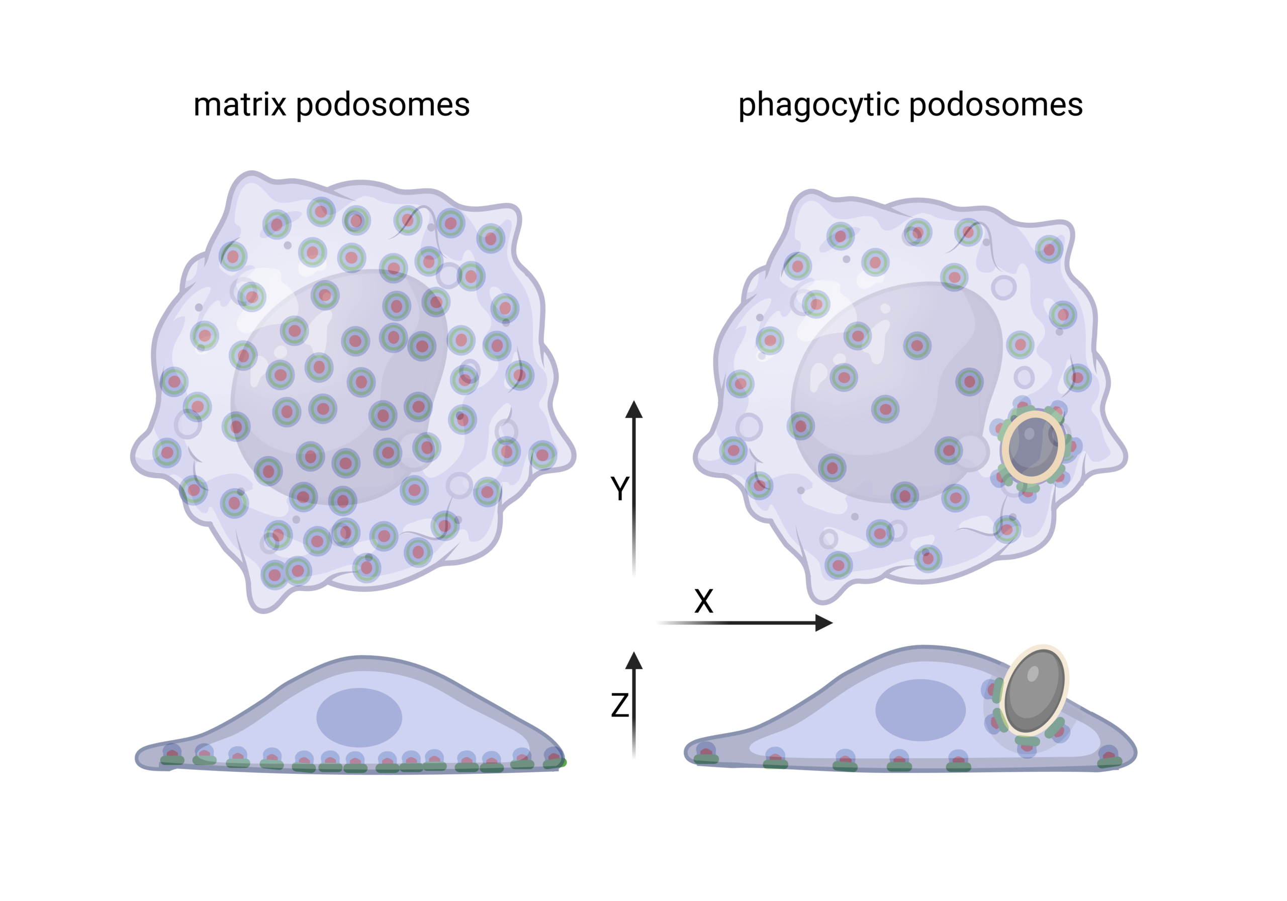
Figure 1: Comparison of ventral and phagocytic podosomes. Adapted from Barcelona and Linder, 2023.
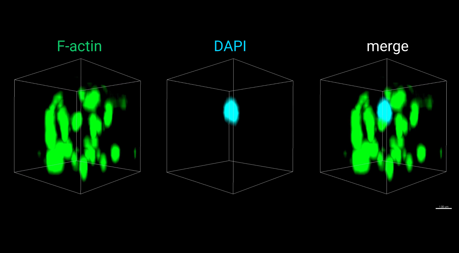
3D-reconstruction of phagocytic podosomes of C. auris. Primary human macrophages were infected with
C. auris cells and stained for F actin to visualize podosomes (green) and DAPI to visualize yeast DNA (blue). Scale bars, 1 μm. Adapted from Sopelniak et al., 2026.
|
We have now characterized phagocytic podosomes as a specialized podosomal subtype formed during the uptake of Candida auris, an emerging pathogen, by primary human macrophages. Hereby, we identify C. auris as the first pathophysiologically relevant target whose internalization involves the formation of phagocytic podosomes. During phagocytic cup formation around Candida cells, phagocytic podosomes share identical architecture to ventral podosomes, including a branched F-actin core, adhesion proteins arranged in a ring structure, a cap structure on top of the actin-rich core containing F-actin regulators, a receptor-rich base, and connecting cables that interlink individual podosomes. |
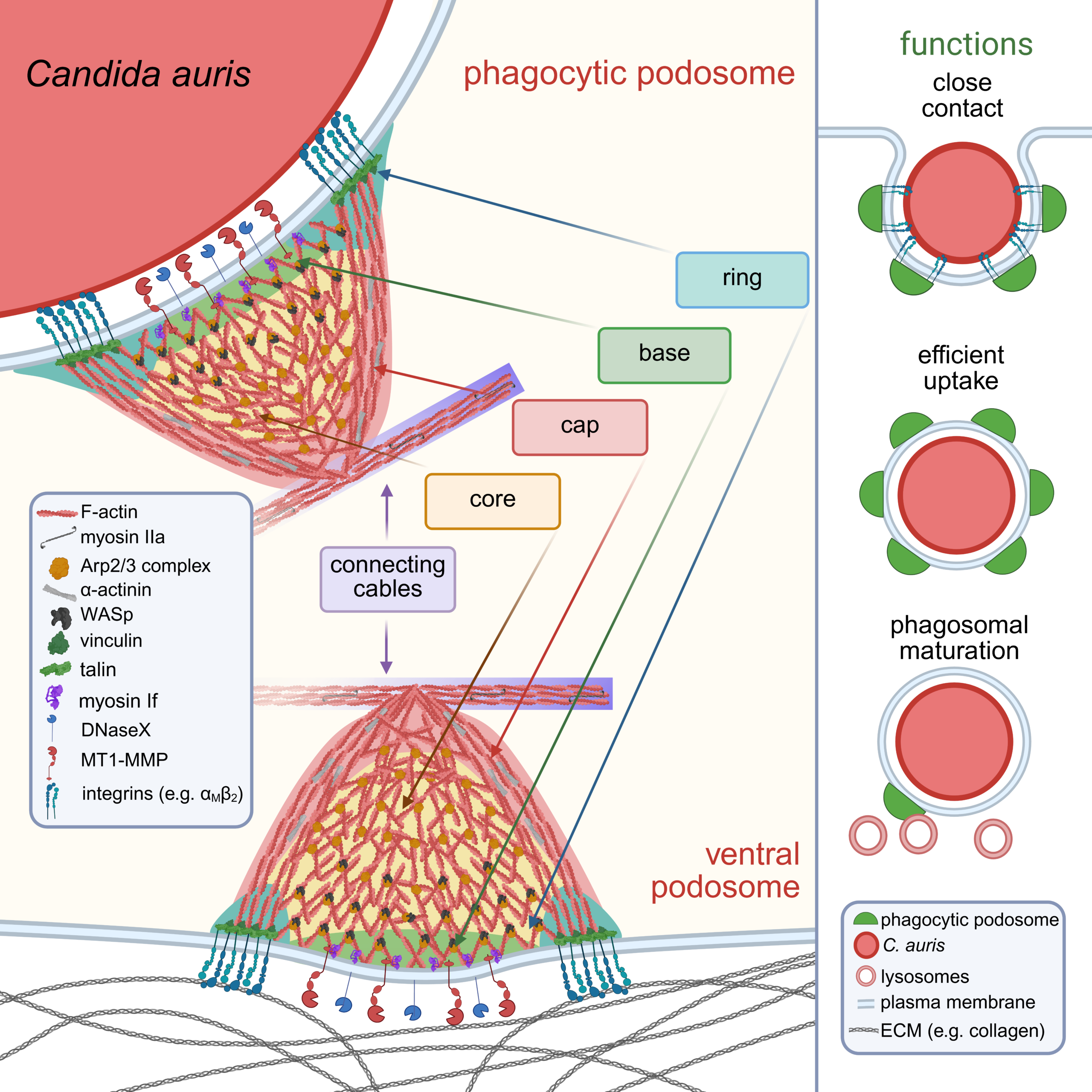
Phagocytic podosomes enable efficient uptake of C. auris. Phagocytic podosomes share identical components as ventral podosomes. Their dynamic organization promotes close contact with the phagocytic target, supports efficient internalization and enhances phagosomal maturation.
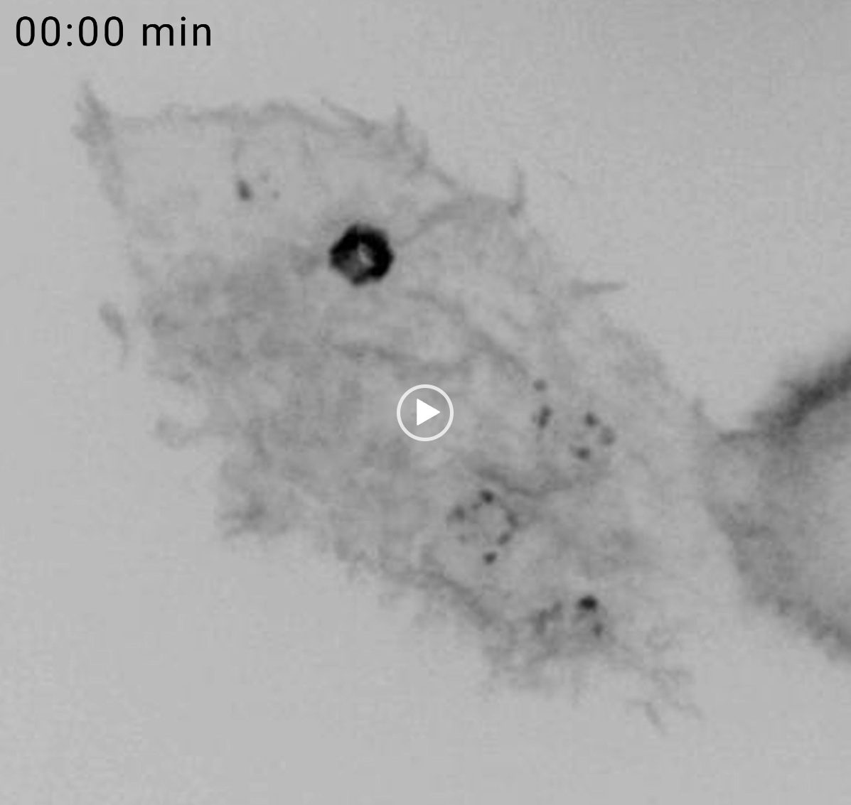
Tracking of phagocytic podosomes during uptake of C. auris. Live-cell imaging of a primary human macrophage expressing LifeAct GFP (grey) and infected with multiple C. auris cells, MOI:50. Maximum projection of optical z-stack with the subtraction of the adhesive plane containing ventral podosomes. Adapted from Sopelniak et al., 2026.
|
Phagocytic podosomes form during the early uptake stage and have a shorter lifespan than their ventral counterparts. Their formation promotes close contact between the macrophage and the fungal cell and is associated with enhanced phagosomal maturation and acidification. This previously uncharacterized mechanism of C. auris phagocytosis suggests that phagocytic podosomes link pathogen recognition, adhesion, and mechanical force transduction, enabling phagosome maturation. Finally, these results also challenge the dogma of a generally uniform and continuous F-actin network in the phagocytic cup. It is likely that phagocytic podosomes are formed during the uptake of comparably large and rigid particles and thus represent an adaptation of the phagocytic cytoskeleton that enables uptake of these difficult targets. Likely through contact with microtubule plus-tip contacts, phagocytic podosomes enable enrichment of lytic enzymes within the phagosomes. Based on this finding, we aim to investigate how the formation of phagocytic podosomes mediates trafficking of lytic enzymes to the nascent phagosome.
Related publications: Sopelniak K, Batlouni R, Sun Q, Cervero P, Linder S. (2026) Linder S, Barcelona B (2023) |